Volume tomography

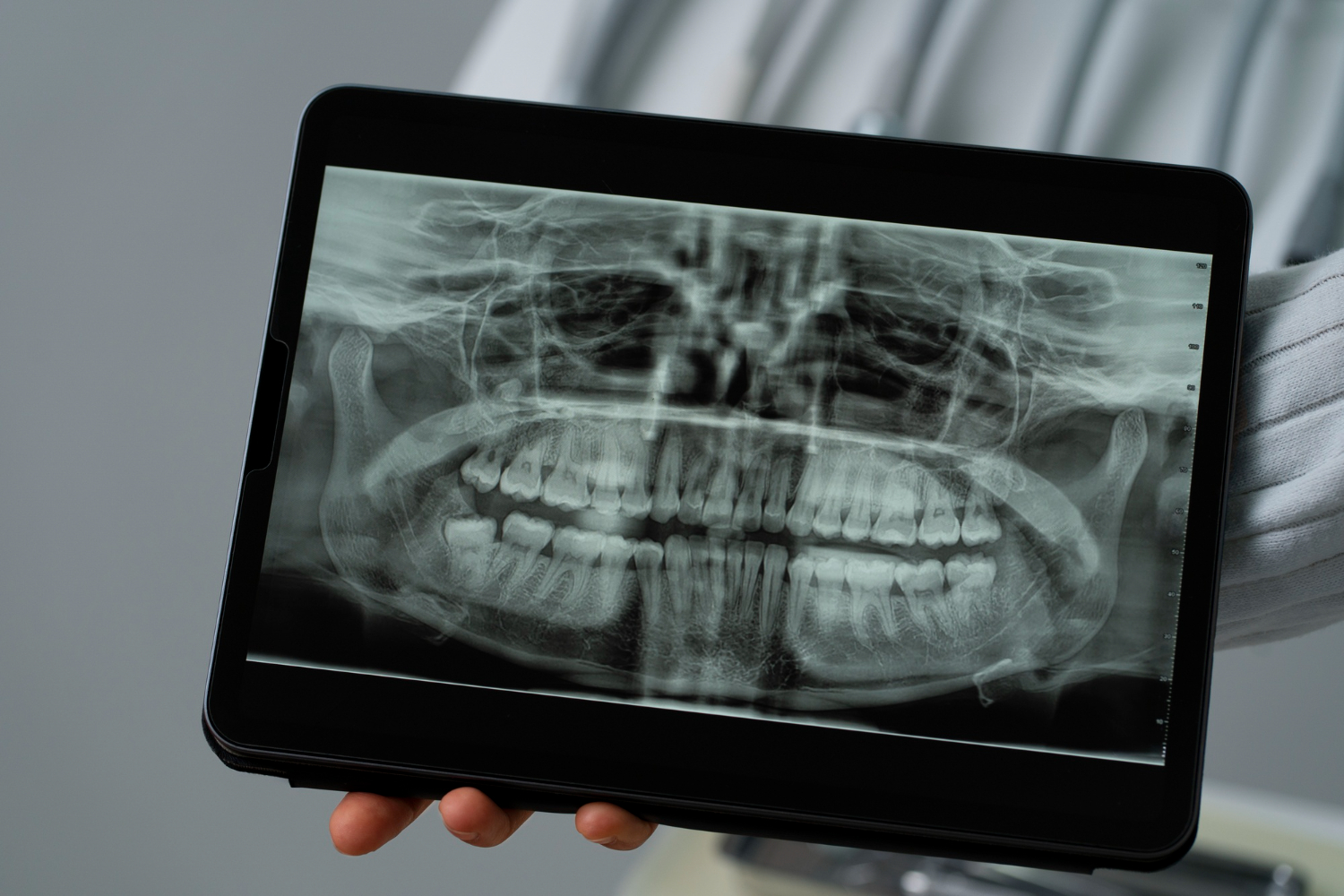
Digital volume tomography (DVT) is a 3D tomography procedure. With this technology we can create precise and detailed 3D images of teeth, jaw bones and surrounding structures. This enables us to make an exact diagnosis and plan treatments.

How does DVT work?

In digital volume tomography, a special X-ray machine rotates once around your head and creates individual images of your mouth and jaw from different angles. These are then combined and assembled into a precise 3D model.

This procedure enables us to:
  • determine the exact position of teeth and their location in the jaw,
  • analyse the course of nerves and blood vessels,
  • detect changes such as cysts, inflammation or bone defects,
  • plan the optimal position for implants.
Good to know: We will discuss with you in advance whether a DVT makes sense in your case and advise you in detail.

How does DVT work?

1.

For the DVT, you stand or sit next to the DVT device. Your chin and forehead are stabilized so that they do not move during the images.

2.

The device then rotates around your head and takes several individual images of your teeth and jaw from different angles.

3.

The individual images are then processed by special software into a detailed 3D model. This model forms the basis for successful diagnosis and treatment.

What are your benefits from a DVT?

  • Precise diagnoses: We can see the smallest details, e.g. the course of roots or the exact position of wisdom teeth.
  • Safety: The technology works with a comparatively low radiation exposure, even though it delivers such detailed images.
  • Better planning: With a DVT we can plan implants and complex procedures precisely and carry them out safely, without any unpleasant surprises.
  • Soft tissue and bone analysis: In addition to teeth, the condition of the surrounding tissue, such as the maxillary sinus, can also be assessed.
Are you suffering from gaps between your teeth, toothache or are you due for root canal treatment? Perhaps there is a suspicion of cysts, bone defects or a complex wisdom tooth position? With digital volume tomography (DVT) we can precisely analyze your problems and plan the optimal treatment.

FAQ

  • Normal X-ray image: Only gives a two-dimensional view – like a photo.
  •  DVT: Shows the structures in 3D, including details that could remain hidden in a flat image.
For dentistry, DVT is usually the better choice: It provides precise 3D images of teeth and jaws with less radiation exposure than a CT and is ideal for diagnosis and treatment planning.
Usually 10-20 seconds.
The radiation exposure during a DVT is comparatively lower and often lower than with conventional CT scans.
A DVT is often used to plan implants, to assess wisdom teeth or to diagnose jaw diseases.
DVT is a safe procedure with minimal radiation exposure. Side effects are rare.
However, you should avoid placing any metal objects around your head and neck.
DVT is suitable for most patients. However, it is only used in urgent cases in pregnant women.